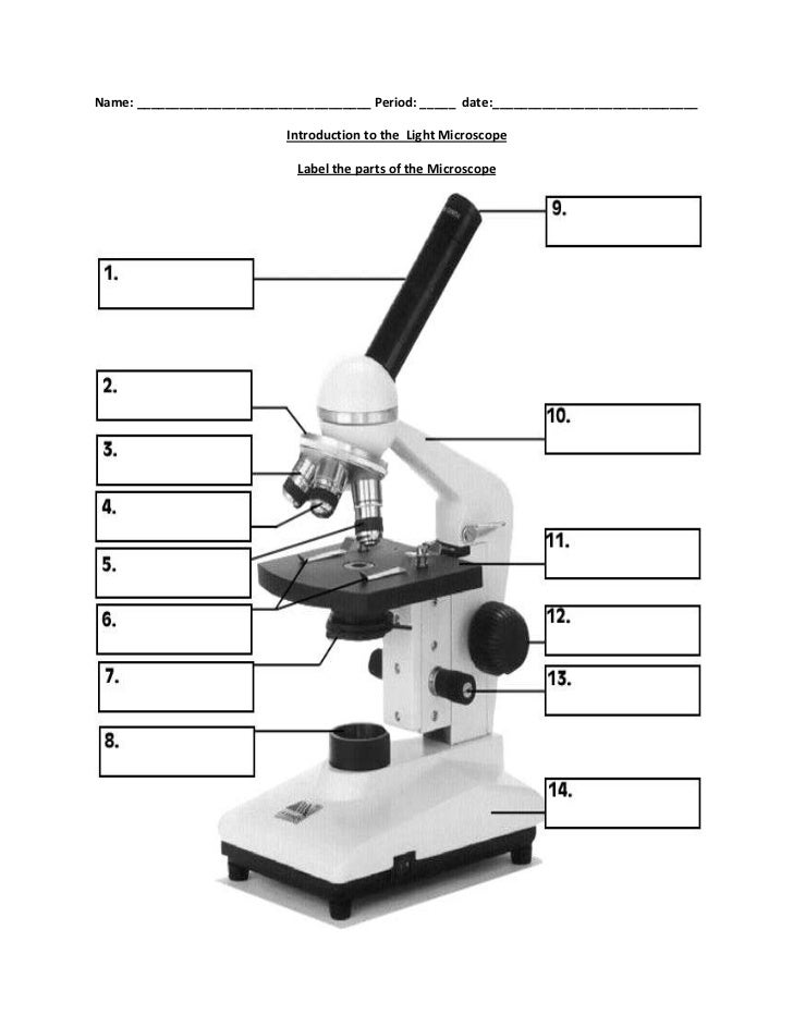
42 labels of a microscope and functions
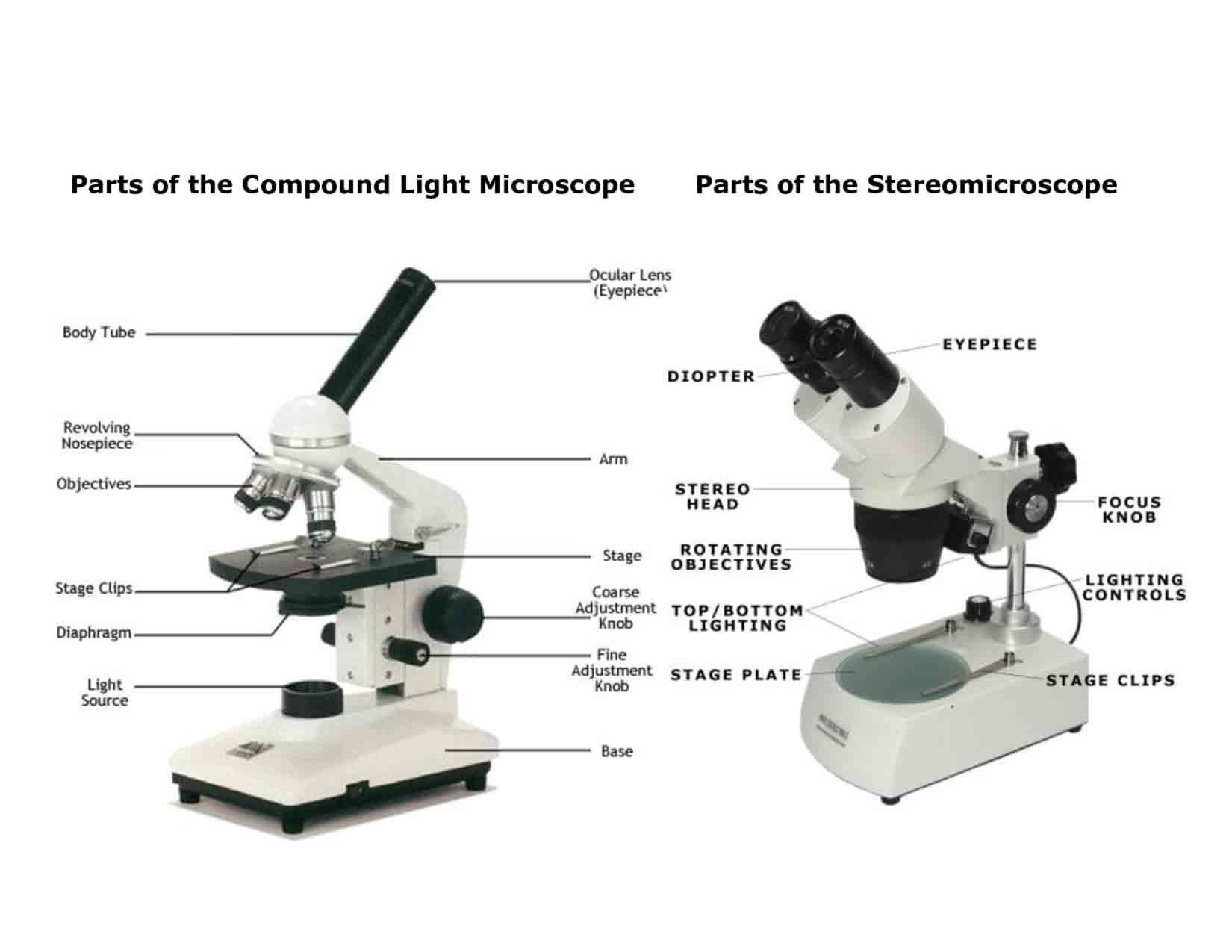
Parts of Stereo Microscope (Dissecting microscope) - labeled diagram ... Stereo microscopes (also called Dissecting microscope) are branched out from other light microscopes for the application of viewing "3D" objects. These include substantial specimens, such as insects, feathers, leaves, rocks, sand grains, gems, coins, and stamps, etc. Functionally, a stereo microscope is like a powerful magnifying glass. Microscope With Labeled Parts and Functions - 24 Hours Of Biology Three are 3 or 4 objective lenses on a microscope .These are the some main lenses that are used for specimen visualization. They have a magnification power of 40x-100X. There are about 1-4 objectives lenses attached to one microscope, in which some are front-facing and others are opposite-facing. Lenses have differing magnification Capacities.
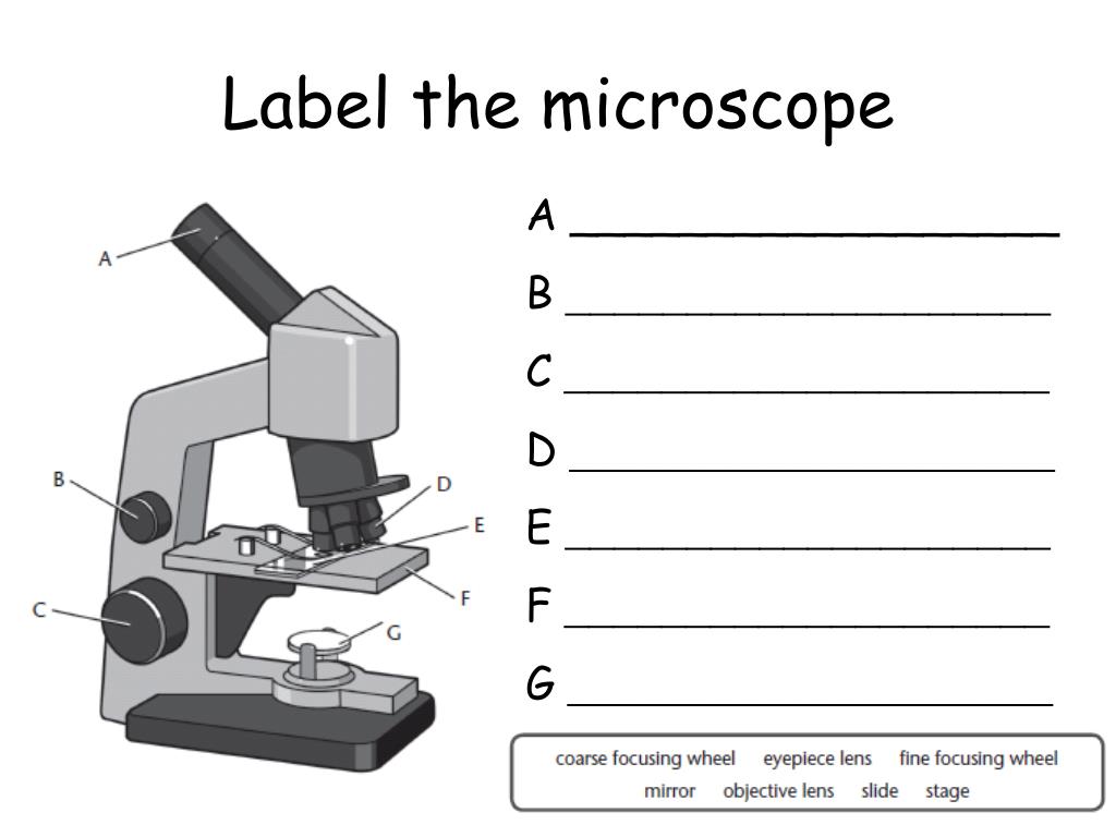
Label the microscope — Science Learning Hub Use this interactive to identify and label the main parts of a microscope. Drag and drop the text labels onto the microscope diagram. stage high-power objective light source eye piece lens base coarse focus adjustment fine focus adjustment diaphragm or iris Download Exercise Tweet
Labels of a microscope and functions
Microscope: Parts Of A Microscope With Functions And Labeled Diagram. Q. Define a Microscope. Ans. Microscopes are instruments that are used in science laboratories, to visualize very minute objects such as cells, and microorganisms, giving a contrasting image, that is magnified. Q. State functions of a microscope. Ans. A microscope is usually used for the study of microscopic algae, fungi, and biological specimens. Compound Microscope Parts - Labeled Diagram and their Functions Basically, compound microscopes generate magnified images through an aligned pair of the objective lens and the ocular lens. In contrast, "simple microscopes" have only one convex lens and function more like glass magnifiers. [In this figure] Two "antique" microscopes played significant roles in the history of biology. Parts of a microscope with functions and labeled diagram - Microbe Notes Optical parts of a microscope and their functions The optical parts of the microscope are used to view, magnify, and produce an image from a specimen placed on a slide. These parts include: Eyepiece - also known as the ocular. This is the part used to look through the microscope. Its found at the top of the microscope.
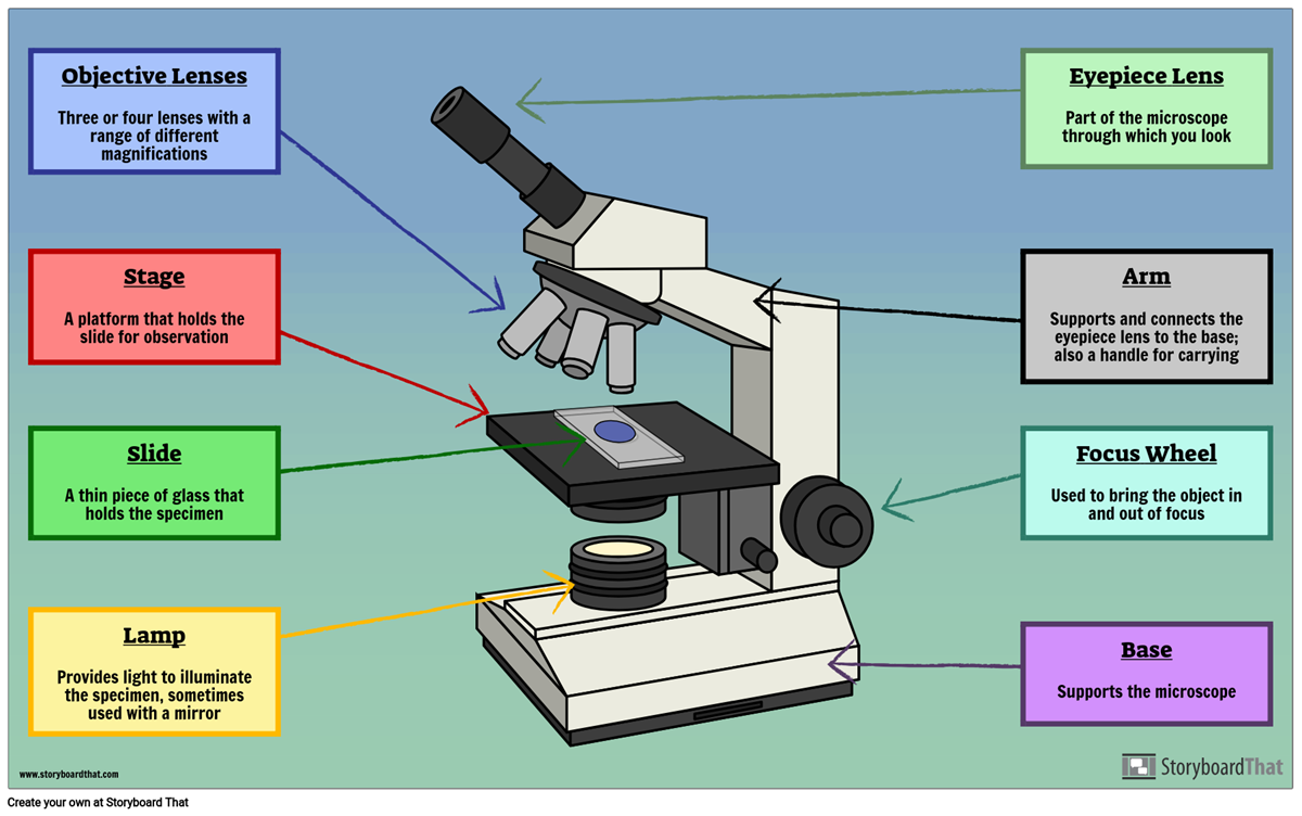
Labels of a microscope and functions. Your liver is essential to your life. The Canadian Liver Foundation This is a myth. Jaundice can be an early warning sign of liver disease. Many babies have “newborn jaundice” lasting three to five days after birth because their liver is not yet fully developed, however, jaundice that does not clear up after 14 days of life, dark urine and/or pale stools, an enlarged abdomen and vomiting are signs that your baby should be seen by his or … Introduction to three-dimensional image processing Introduction to three-dimensional image processing¶. Images are represented as numpy arrays. A single-channel, or grayscale, image is a 2D matrix of pixel intensities of shape (row, column).We can construct a 3D volume as a series of 2D planes, giving 3D images the shape (plane, row, column).Multichannel data adds a channel dimension in the final position … Parts of Stereo Microscope (Dissecting microscope) – labeled … Unlike a compound microscope that offers a flat image, stereo microscopes give the viewer a 3-dimensional image that you can see the texture of a larger specimen. [In this image] Examples of Stereo & Dissecting microscopes. Major microscope brands (Zeiss, Olympus, Nikon, Amscope, Omano, Leica …) all produce stereomicroscopes. Parts of a Microscope Labeling Activity - Storyboard That Create a poster that labels the parts of a microscope and includes descriptions of what each part does. Click "Start Assignment". Use a landscape poster layout (large or small). Search for a diagram of a microscope. Using arrows and textables label each part of the microscope and describe its function.
Compound Microscope Parts, Functions, and Labeled Diagram Compound Microscope Definitions for Labels. Eyepiece (ocular lens) with or without Pointer: The part that is looked through at the top of the compound microscope. Eyepieces typically have a magnification between 5x & 30x. Monocular or Binocular Head: Structural support that holds & connects the eyepieces to the objective lenses. ch 8 mastering biology Flashcards | Quizlet Drag the pink labels onto the pink targets to identify the two main phases of the cell cycle. ... the cell carries out its normal functions and the chromosomes are thinly spread out throughout the nucleus. interphase. Part E Looking through a light microscope at a dividing cell, you see two separate groups of chromosomes on opposite ends of the ... The Parts of a Microscope (Labeled) Printable - TeacherVision The Parts of a Microscope (Labeled) Printable. Download. Add to Favorites. Share. This diagram labels and explains the function of each part of a microscope. Use this printable as a handout or transparency to help prepare students for working with laboratory equipment. Grade: Microscope Types (with labeled diagrams) and Functions Simple microscope labeled diagram Simple microscope functions It is used in industrial applications like: Watchmakers to assemble watches Cloth industry to count the number of threads or fibers in a cloth Jewelers to examine the finer parts of jewelry Miniature artists to examine and build their work Also used to inspect finer details on products
Dissecting Microscope Parts And Functions. All You Need To Know The stand/arm is the spine of the dissecting microscope because its function is to provide support for the head while at the same time connecting the head to the microscope base. The microscope's design will determine if the stand is hollow, an immobile arm or cylindrical rod. To light the specimen from above the power cable will extend from ... Microscope, Microscope Parts, Labeled Diagram, and Functions Microscopes magnify or enlarge small objects such as cells, microbes, bacteria, viruses, microorganisms etc. at a viewable scale for examination and analysis. Microscopes consist of one or more magnification lenses to enlarge the image of the microscopic objects placed in the focal plane. A Study of the Microscope and its Functions With a Labeled Diagram ... A Study of the Microscope and its Functions With a Labeled Diagram To better understand the structure and function of a microscope, we need to take a look at the labeled microscope diagrams of the compound and electron microscope. These diagrams clearly explain the functioning of the microscopes along with their respective parts. Microscope Parts and Functions Flashcards | Quizlet Magnifies image 40X found on the nosepiece. Base. Support/bottom of the microscope, used to carry the microscope. Light Source. Provides light to enable us to see the specimen on the slide. Arm. Used in order to carry the microscope. Coarse Adjustment. moves the stage up or down a lot, used first when viewing the slide.
Microscope Parts and Functions First, the purpose of a microscope is to magnify a small object or to magnify the fine details of a larger object in order to examine minute specimens that cannot be seen by the naked eye. Here are the important compound microscope parts... Eyepiece: The lens the viewer looks through to see the specimen.
Microscope Parts & Functions - AmScope Microscope Parts and Functions Invented by a Dutch spectacle maker in the late 16th century, compound light microscopes use two sets of lenses to magnify images for study and observation. The first set of lenses are the oculars, or eyepieces, that the viewer looks into; the second set of lenses are the objectives, which are closest to the specimen.
Label the microscope — Science Learning Hub Jun 08, 2018 · All microscopes share features in common. In this interactive, you can label the different parts of a microscope. Use this with the Microscope parts activity to help students identify and label the main parts of a microscope and then describe their functions.. Drag and drop the text labels onto the microscope diagram. If you want to redo an answer, click on the …
22 Parts Of a Microscope With Their Function And Labeled Diagram Invented by a Dutch spectacle maker in the late 16th century, light microscopes use lenses and light to magnify images. Generally a microscope works on the basis of resolution and magnification. The magnifying power of a microscope is an expression of the number of times the object being examined appears to be enlarged and is a dimensionless ratio.
Microscope Parts, Function, & Labeled Diagram - slidingmotion Microscope parts labeled diagram gives us all the information about its parts and their position in the microscope. Microscope Parts Labeled Diagram The principle of the Microscope gives you an exact reason to use it. It works on the 3 principles. Magnification Resolving Power Numerical Aperture. Parts of Microscope Head Base Arm Eyepiece Lens
Electron microscope - Wikipedia An electron microscope is a microscope that uses a beam of accelerated electrons as a source of illumination. As the wavelength of an electron can be up to 100,000 times shorter than that of visible light photons, electron microscopes have a higher resolving power than light microscopes and can reveal the structure of smaller objects.. Electron microscopes use shaped magnetic …
Temporal analysis of enhancers during mouse cerebellar ... - eLife Aug 09, 2022 · Temporal analysis of enhancers during mouse cerebellar development reveals dynamic and novel regulatory functions. Miguel Ramirez ... The datasets collected at these ages and the downstream analyses are shown in the flow chart. Labels: NE ... Analysis and photomicroscopy were performed using a Zeiss Axiovert 200 M microscope with the Axiocam ...
Parts of the Microscope with Labeling (also Free Printouts) Let us take a look at the different parts of microscopes and their respective functions. 1. Eyepiece it is the topmost part of the microscope. Through the eyepiece, you can visualize the object being studied. Its magnification capacity ranges between 10 and 15 times. 2. Body tube/Head It is the structure that connects the eyepiece to the lenses.
Robert Hooke - Biography, Facts and Pictures - Famous Scientists Robert Hooke was a Renaissance Man - a jack of all trades, and a master of many. He wrote one of the most significant scientific books ever written, Micrographia, and made contributions to human knowledge spanning Architecture, Astronomy, Biology, Chemistry, Physics, Surveying & Map Making, and the design and construction of scientific instruments.
Microscope Parts and Functions - YouTube This video goes along with your microscope parts and function worksheet (Microscope Lab)
Fluorescent Nanodiamonds for HeLa Cell Drug Delivery Aug 24, 2022 · Besides drug delivery, the optical properties and photostability of fluorescent nanodiamonds extend their applications in long-term optical imaging. Moreover, due to the visibility of fluorescent nanodiamonds through different imaging techniques, they are also applied as labels for correlative microscopy.
Confocal Microscopy - an overview | ScienceDirect Topics A confocal microscope was invented in 1951 by Marvin Minsky, a postdoctoral fellow at Harvard University studying neural networks in living brain (Minsky, 1988).In 1957, Minsky patented the concept of confocal imaging, the illumination and detection of a single diffraction-limited spot in a specimen (Fig. 1A).In the transmission configuration, the condenser is replaced with a second …
Parts of a microscope with functions and labeled diagram - Microbe Notes Optical parts of a microscope and their functions The optical parts of the microscope are used to view, magnify, and produce an image from a specimen placed on a slide. These parts include: Eyepiece - also known as the ocular. This is the part used to look through the microscope. Its found at the top of the microscope.
Compound Microscope Parts - Labeled Diagram and their Functions Basically, compound microscopes generate magnified images through an aligned pair of the objective lens and the ocular lens. In contrast, "simple microscopes" have only one convex lens and function more like glass magnifiers. [In this figure] Two "antique" microscopes played significant roles in the history of biology.
Microscope: Parts Of A Microscope With Functions And Labeled Diagram. Q. Define a Microscope. Ans. Microscopes are instruments that are used in science laboratories, to visualize very minute objects such as cells, and microorganisms, giving a contrasting image, that is magnified. Q. State functions of a microscope. Ans. A microscope is usually used for the study of microscopic algae, fungi, and biological specimens.






Post a Comment for "42 labels of a microscope and functions"